

Reaction Disclaimer

On the screen, two yellow-coloured cells are visible, positioned diagonally above one another. Suddenly, a red-purple wave sweeps through the cells from the lower right. This is the effect of a detergent that Joachim Goedhart applies so that all the calcium from the medium enters the mitochondria. We observe this effect thanks to a new calcium biosensor — one that, remarkably enough, does not measure the intensity of the fluorescence, but rather the lifetime of the fluorophore. More on that shortly.
The use of fluorescent proteins and biosensors is becoming increasingly commonplace — not only to visualise where a particular protein is located, but also to investigate what it does. Developing these tools is a field in its own right, in which knowledge, expertise and luck all play a role. Since the discovery of green fluorescent protein (GFP), researchers have learnt a great deal: about which modifications make the protein brighter or cause it to fluoresce in a different colour, and how to couple the protein to a biosensor. ‘But I actually think we still have a surprisingly limited understanding of precisely how these things make themselves fluorescent,’ says Dorus Gadella, Professor of Molecular Cytology at the University of Amsterdam, whose group develops new fluorescent proteins and biosensors.
Using mutations, they introduce changes at sites they know or suspect to influence fluorescence, after which they screen for desired outcomes. ‘Where you ultimately end up depends on which screening methods you use for that purpose,’ says Joachim Goedhart, Assistant Professor of Molecular Cytology. ‘You can screen for colour variants, or for photostability. And one of the original contributions of our laboratory is that we started looking at lifetime.’
‘We still have a surprisingly limited understanding of precisely how these things make themselves fluorescent’
Dorus Gadella
Gadella explains this using a 3D-printed model of mTurquoise2, a blue fluorescent protein derived from a bioluminescent jellyfish. The source of the fluorescence is a fluorophore consisting of three amino acids: serine, tryptophan and glycine. The remainder of the protein is folded around it, forming what is known as a barrel. This ‘barrel’ consists of beta-sheets and holds the fluorophore in place. By absorbing photons of a specific wavelength, the electrons in the fluorophore begin to vibrate, breaking contact with the surrounding barrel. The absorbed energy can then be released by the fluorophore either by emitting photons of slightly lower energy, or by flipping over.
‘You can essentially attach or connect all manner of things to those new termini’
Joachim Goedhart
‘And what we do with lifetime is look at how long the fluorophore is able to hold onto that excited energy before it fluoresces. For a fluorescent protein, that is a fixed value — dependent solely on the chemistry of the chromophores,’ says Gadella. This makes it independent of the microscopy setup, in contrast to fluorescence intensity, which researchers typically use. That intensity is also dependent on the number of molecules, the laser used, and the quality of the detector.
So much for the fluorescent proteins. For biosensors, Goedhart substantially remodels the fluorescent protein. He joins the original termini of the protein together and creates two new termini in the barrel, close to the point that holds the fluorophore in place. These new termini are the essence of fluorescent biosensors. ‘You can essentially attach or connect all manner of things to those new termini,’ says Goedhart. In the case of a calcium sensor, these are two domains that bind more tightly to one another as calcium concentrations increase. This interaction causes the barrel to shift slightly, thereby holding the fluorophore either more or less firmly in place.
‘This has been done very frequently with GFP, and we were keen to explore whether, instead of a simple intensity change, you could also bring about a lifetime change — because lifetime is so robust,’ says Goedhart. This resulted in the turquoise and green Ca-FLITS biosensors, developed in collaboration with the now-graduated Franka van der Linden. The key advantage is that the lifetime of the Ca-FLITS biosensors correlates one-to-one with the calcium concentration, meaning researchers no longer need to go through endless calibration every time they use the sensor.
‘You can really see what’s happening, just with your own eyes’
Jaap van Buul
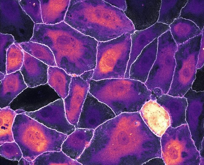
During the development of new biosensors and fluorescent proteins, Gadella’s group uses simplified systems that are straightforward to screen and work with. ‘So you have a very simple cell system, which doesn’t really have much to do with the biology,’ says Jaap van Buul, Professor of Vascular Cell Biology at the University of Amsterdam. ‘But the difficulty lies precisely in sometimes finding a suitable biological system to go with it — and that’s where we come in.’ He investigates how leucocytes locate an infection on the outer surface of a blood vessel wall in order to squeeze through at exactly that point. In doing so, he collaborates closely with the molecular cytology group. According to Van Buul, the advantage of the probes is that ‘you can really see what’s happening, just with your own eyes.’
One of the biosensors Van Buul uses reveals protein activation at highly localised sites within the cell. ‘We can then see, for example, that the protein becomes active only at a specific location, and nowhere else. And it turns out that this is precisely where the leucocyte passes through.’ For Van Buul, the collaboration with the molecular cytology group has become indispensable, and Gadella and Goedhart likewise draw their motivation from it — the drive to keep tinkering with those molecules to make them ever better and brighter. Before long, Werner van der Meer, a PhD candidate in Van Buul’s group, will begin working with the new Ca-FLITS biosensor, with the aim of determining whether there is a local calcium signal that corresponds to the site where the leucocyte passes through the blood vessel wall. For Gadella and Goedhart, this will be the test of whether Ca-FLITS genuinely yields new insights in a complex biological system.


Every Wednesday in your inbox: the latest chemistry and life sciences news from the Netherlands and Flanders, plus analyses, background stories and more!


No comments yet
Only registered users can post comments on this article.
Log in or register to comment.